 |
Case Report
Management of an intercostal-pulmonary artery fistula in the setting of massive hemoptysis
1 MD, Resident, Department of Surgery, Riverside Methodist Hospital, Columbus, OH, USA
2 MD, Cardiothoracic Surgeon, Department of Surgery, Riverside Methodist Hospital, Columbus, OH, USA
Address correspondence to:
Danielle Hery
MD, OhioHealth Riverside Methodist Hospital, 3535 Olentangy River Road, Columbus, OH 43214,
USA
Message to Corresponding Author
Article ID: 100112Z06EG2022
Access full text article on other devices

Access PDF of article on other devices


How to cite this article
Ghazoul E, Hery D, Wu J, Kim W. Management of an intercostal-pulmonary artery fistula in the setting of massive hemoptysis. Case Rep Int 2022;11(2):20–24.ABSTRACT
Introduction: Intercostal artery to pulmonary artery fistulas is exceptionally rare. The few cases reported in the literature are often found in asymptomatic patients, but this physiology can result in massive hemoptysis and subsequent respiratory failure secondary to aspiration.
Case Report: A male in his 40s presented to our tertiary community hospital with massive hemoptysis and a known diagnosis of Mycobacterium szulgai; his past surgical history was pertinent for a coronary artery bypass with chest tube placement four years prior to his presentation and for remote pneumothoraces bilaterally. Angiography identified a fistulous connection between multiple intercostal artery trunks to a right upper lobe pulmonary artery that was coil embolized, successfully resolving his hemoptysis.
Conclusion: This case report investigates a potential complication of those in an active inflammatory state, such as a mycobacterium infection, and the opportunity for minimally invasive treatment for those with massive hemoptysis.
Keywords: Fistula, Hemoptysis, Mycobacterium szulgai, Pulmonary artery
INTRODUCTION
Intercostal arteries to pulmonary artery fistulas are exceptionally rare. They are often asymptomatic and found incidentally with a heart murmur or rib notching on a chest X-ray. There is little knowledge on their treatment as examples are rare [1]. In general, fistulas are often caused by trauma or surgical interventions and there have been reports of associations with inflammation or infection. In a scenario with both trauma and infection, it is difficult to assess the initial insult, but potentially both factors perpetuated an ongoing fistula causing massive hemoptysis. We review a potential complication of an active inflammatory state, the importance of early involvement of the cardiothoracic team and interventional radiology, and a successful minimally invasive approach to management at our institution.
CASE REPORT
A male in his mid-40s presented to our tertiary care hospital with hemoptysis. He had a known diagnosis of Mycobacterium szulgai and was being treated by infectious disease as an outpatient. His past medical history was significant for 15 pack-years of smoking, marijuana use, chronic obstructive pulmonary disease (COPD), a prior three-vessel coronary artery bypass graft (CABG) four years prior to presentation, and bilateral pneumothoraces managed with thoracostomy tubes many years prior to his CABG. Nine months prior to his presentation, he began having increased sputum production and mild hemoptysis with a chest radiograph concerning for a right upper lobe inflammatory process. A computed tomography (CT) image of the chest was obtained showing a partially cavitary infiltrate in the right upper lobe. He underwent a bronchoscopy and transbronchial drainage of the infiltrate, which grew group B streptococcus, Pseudomonas, and a slow growing M. szulgai. He was treated with Augmentin and ciprofloxacin for seven days and was referred to an outpatient infectious disease team on discharge. There he was treated with rifabutin, azithromycin, and ethambutol. His azithromycin was changed to clarithromycin for tolerability four months prior to presentation at our institution.
The patient presented to an outlying hospital with increasing bright red hemoptysis for the past two weeks. He received tranexamic acid and blood transfusions after another episode of sub-massive hemoptysis, then was transferred to the medical intensive care unit at our facility with a thoracic surgical evaluation.
Physical exam was notable for a frail male, appearing older than his stated age. Lungs were clear to auscultation, and no bruits palpable masses were found.
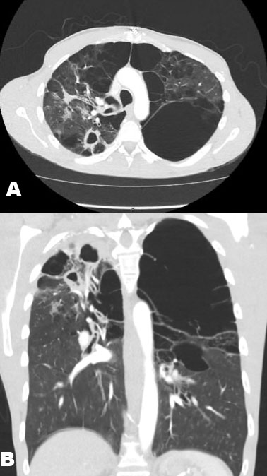
A CT angiogram of the chest was obtained, which showed stable cavitary lesions, the two largest sized at 2.7 and 2.4 cm, in the right upper lobe, significant bronchiectasis, massive bullous emphysema, and parenchymal ground-glass opacities (Figure 1A and Figure 1B). There was no evidence of arterial extravasation. At the time of thoracic surgical evaluation, the hemoglobin was 14.4 g/dL.
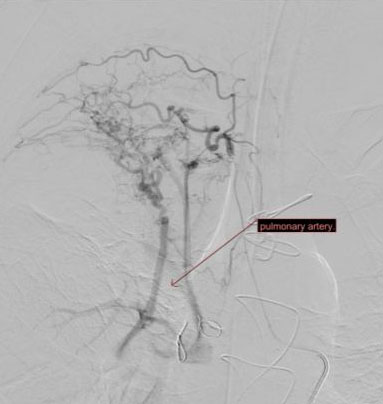
The patient was hemodynamically stable and electively intubated for bronchoscopy and interventional radiology evaluation. Bronchoscopy revealed old blood without signs of source or active bleed. The patient remained hemodynamically stable throughout. On hospital day 1, the patient was taken by interventional radiology for an angiogram and possible embolization. The angiogram revealed a hypertrophied right 3rd, 4th, and 5th intercostal trunk arteries that had a fistulous communication via multiple vessels to the right upper lobe pulmonary artery (Figure 2). Particle embolization was attempted, but the fistula was too large. Coil embolization was then performed, successfully occluding the fistulous connection. The patient did well post-procedure. He had no further episodes of significant hemoptysis, and his hemoglobin remained stable. He was extubated post-procedure day 1 and was discharged on hospital day 4.
DISCUSSION
Vascular fistulas have been known to develop as a result of surgical interventions on the thoracic cavity, often multiple years after the incident. There have been case reports describing tube thoracostomies causing fistulas of the chest walls [2],[3],[4]. One of the first reported cases of thoracostomy-induced systemic to pulmonary arterial fistula was in 1965 and involved an Air Force officer who had had a tube thoracostomy for a spontaneous pneumothorax. He presented with a bruit over the right chest wall near where the tube thoracostomy had been located [4]. In the case reported by Hirsch, the patient only manifested his fistula with a precordial murmur heart on auscultation. There have been cases reported in literature prior to these instances, but they were largely deemed to be congenital in origin [3],[4]. One review of the literature demonstrated etiologies spanning from neoplastic, to congenital, and inflammatory, with few falling within the traumatic origin [3]. For instance, another case of pulmonary to intercostal artery fistula described a completely asymptomatic teenager found to have rib-notching on a routine screening radiograph; he had no prior traumas or thoracic procedures. It can be posited that those found in the younger patient population without thoracic surgical or trauma history can be attributed to congenital cases [1]. The patient described in the presented case at our institution had not only an open cardiac procedure, but bilateral chest tubes years prior his presentation to our hospital; the trauma from the placement of these or the cardiac surgery itself is one of the most likely etiologies of his fistula; however, his chronic infection with M. szulgai also lent itself as another risk factor for systemic to pulmonary arterial fistula formation.
Mycobacterium szulgai accounts for less than 0.2% of atypical or non-tuberculosis mycobacterium. It is slow growing and most commonly presents with pulmonary infection and can act very similar to active pulmonary tuberculosis. It is generally diagnosed in immune-compromised individuals or those with COPD and smoking history [5]. Because of the low prevalence, there is not a standard treatment regimen. However, case studies have shown it is susceptible to multi-drug anti-tuberculosis regimens [6]. There have been no reported complications of vascular fistulas from M. szulgai specifically, but there is literature detailing systemic to pulmonary artery fistula after a pulmonary abscess was drained growing Actinomyces israeli [7]. Another case described in the literature of a systemic to pulmonary communication involved Mycobacterium tuberculosis, which led to a patient presenting with high cardiac output heart failure. This case demonstrates another instance in which no prior thoracic surgical interventions or trauma had been performed [8]. It appears that these fistulas can occur in chronic and granulomatous conditions. The inflammation from the disease process along with the lung abscess drainage.
The formation of an intercostal artery to pulmonary artery fistula is largely thought to be due to an insidious inflammatory process from his pneumonia and previous manipulation of his chest wall. There is a potential that the prior chest tubes could have precipitated the neovascular formation of the fistula years before his presentation. The active and chronic inflammation from the nontuberculous Mycobacterium causing an increase in size, thus propagating his hemoptysis.
Treatment of these fistulas has evolved over time. In the first recorded case of treatment of a traumatic pulmonary to systemic arterial fistula, open thoracotomy, and electrocautery were used to control and ligate the aberrant vasculature [4]. Lobectomy was preferred in the majority of the congenital cases described prior to that time, though open ligation and division of the feeding vessel was used as well to a lesser extent. One of the first recorded cases of using transcatheter embolization of the aberrant vasculature was in 1983 [3]. Minimally invasive endovascular approaches have been described in the literature subsequently as reliable methods of fistula control and cure [1],[8],[9],[10],[11],[12],[13].
Due to the rarity of systemic-to-pulmonary arterial fistulas, there is not a definitive way to manage and treat these fistulas. This minimally invasive approach not only utilizes angiography to diagnose, but in the same procedure, embolization allows the fistula to be closed quickly and within minimal postoperative complications when in comparison to a larger surgical intervention. Follow-up after embolization is necessary, as these embolized sites are able to recanalize over time [11].
CONCLUSION
Intercostal to pulmonary arterial fistulas are extremely rare and are generally asymptomatic. Those in a chronic inflammatory state, as well as history of prior thoracic surgical interventions presenting with hemoptysis should lead the diagnosing team to consider these vascular malformations high on their differentials. Prompt angiography and embolization are effective and minimally invasive approaches to treating these fistulas.
REFERENCE
1.
Cantasdemir M, Kantarci F, Islak C, Kocer N, Saltuk L, Numan F. Transcatheter coil embolization of an intercostal artery to pulmonary artery fistula. Eur Radiol 2002;12(2):454–7. [CrossRef]
[Pubmed]

2.
Coulter TD, Maurer JR, Miller MT, Mehta AC. Chest wall arteriovenous fistula: An unusual complication after chest tube placement. Ann Thorac Surg 1999;67(3):849–50 [CrossRef]
[Pubmed]
3.
Hirsch M, Maroko I, Gueron M, Goleman L. Systemic-pulmonary arteriovenous fistula of traumatic origin: A case report. Cardiovasc Intervent Radiol 1983;6(3):160–3. [CrossRef]
[Pubmed]
4.
Cox PA, Keshishian JM, Blades BB. Traumatic arteriovenous fistula of the chest wall and lung. Secondary to insertion of an intercostal catheter. J Thorac Cardiovasc Surg 1967;54(1):109–12.
[Pubmed]
5.
van Ingen J, Boeree MJ, de Lange WCM, de Haas PEW, Dekhuijzen PNR, van Soolingen D. Clinical relevance of Mycobacterium szulgai in The Netherlands. Clin Infect Dis 2008;46(8):1200–5. [CrossRef]
[Pubmed]
6.
Gido RDS, Wojciechowski AL, Bajwa RP. Pulmonary infection with Mycobacterium szulgai: A case report. SAGE Open Med Case Rep 2019;7:2050313X18823448. [CrossRef]
[Pubmed]
7.
Knoepfli HJ, Friedli B. Systemic-to-pulmonary artery fistula following actinomycosis. Chest 1975;67(4):494–6. [CrossRef]
[Pubmed]
8.
Chino M, Kawaguchi T, Sakai T, Okuno T. Intercostal-to- pulmonary arterial anastomosis, complicated by high-output heart failure: Case report. Angiology 1991;42(3):256–60. [CrossRef]
[Pubmed]
9.
Ferro C, Rossi UG, Bovio G, Seitun S, Rossi GA. Percutaneous transcatheter embolization of a large pulmonary arteriovenous fistula with an Amplatzer vascular plug. Cardiovasc Intervent Radiol 2007;30(2):328–31. [CrossRef]
[Pubmed]
10.
Rossi M, Rebonato A, Greco L, et al. A new device for vascular embolization: Report on case of two pulmonary arteriovenous fistulas embolization using the amplatzer vascular plug. Cardiovasc Intervent Radiol 2006;29(5):902–6. [CrossRef]
[Pubmed]
11.
Cartin-Ceba R, Swanson KL, Krowka MJ. Pulmonary arteriovenous malformations. Chest 2013;144(3):1033–44. [CrossRef]
[Pubmed]
12.
Fu Z, Liang Y, Zhao W, Tian J, Cai F, Zhang X. Safety and efficacy of transcatheter embolization in patients with massive hemoptysis due to intercostal pulmonary venous shunts. Radiol Med 2019;124(7):588–94. [CrossRef]
[Pubmed]
13.
Chilvers ER, Whyte MK, Jackson JE, Allison DJ, Hughes JM. Effect of percutaneous transcatheter embolization on pulmonary function, right-to-left shunt, and arterial oxygenation in patients with pulmonary arteriovenous malformations. Am Rev Respir Dis 1990;142(2):420–5. [CrossRef]
[Pubmed]
SUPPORTING INFORMATION
Author Contributions
Erika Ghazoul - Conception of the work, Design of the work, Acquisition of data, Analysis of data, Drafting the work, Revising the work critically for important intellectual content, Final approval of the version to be published, Agree to be accountable for all aspects of the work in ensuring that questions related to the accuracy or integrity of any part of the work are appropriately investigated and resolved.
Danielle Hery - Conception of the work, Design of the work, Acquisition of data, Analysis of data, Drafting the work, Revising the work critically for important intellectual content, Final approval of the version to be published, Agree to be accountable for all aspects of the work in ensuring that questions related to the accuracy or integrity of any part of the work are appropriately investigated and resolved.
Jin Wu - Conception of the work, Design of the work, Revising the work critically for important intellectual content, Final approval of the version to be published, Agree to be accountable for all aspects of the work in ensuring that questions related to the accuracy or integrity of any part of the work are appropriately investigated and resolved.
Wesley Kim - Conception of the work, Design of the work, Revising the work critically for important intellectual content, Final approval of the version to be published, Agree to be accountable for all aspects of the work in ensuring that questions related to the accuracy or integrity of any part of the work are appropriately investigated and resolved.
Guarantor of SubmissionThe corresponding author is the guarantor of submission.
Source of SupportNone
Consent StatementWritten informed consent was obtained from the patient for publication of this article.
Data AvailabilityAll relevant data are within the paper and its Supporting Information files.
Conflict of InterestAuthors declare no conflict of interest.
Copyright© 2022 Erika Ghazoul et al. This article is distributed under the terms of Creative Commons Attribution License which permits unrestricted use, distribution and reproduction in any medium provided the original author(s) and original publisher are properly credited. Please see the copyright policy on the journal website for more information.


