 |
Case Report
Atypical presentation of a large pneumothorax in a young male: A case report
1 Security Forces Hospital Program, Emergency Department, Riyadh City, Kingdom of Saudi Arabia
2 Emergency Department, Security Forces Hospital, Riyadh City, Kingdom of Saudi Arabia
Address correspondence to:
Yahia Yaseen Akeely
MD, Security Forces Hospital Program, Emergency Department, Riyadh City 11481, P.O. Box 3643,
Kingdom of Saudi Arabia
Message to Corresponding Author
Article ID: 100110Z06YK2022
Access full text article on other devices

Access PDF of article on other devices


How to cite this article
Akeely YY, Alenezy AS, Al Marzouqi SM, Bokhari N, Yousef M. Atypical presentation of a large pneumothorax in a young male: A case report. Case Rep Int 2022;11(2):11–15.ABSTRACT
Introduction: Pneumothorax is a potentially dangerous condition that, if not properly recognized and treated, can have fatal consequences. Chest discomfort or difficulty breathing is common presenting symptoms. We present an atypical presentation for pneumothorax.
Case Report: A 26-year-old male patient presented to the emergency department (ED) with complaints of pain in the right iliac fossa with localized rebound tenderness. The treating physician ordered a computed tomography (CT) abdomen to assess for acute appendicitis. From the abdominal CT scan, the radiologist reported a significant right-sided pneumothorax. The patient was treated with a chest thoracostomy and had an unremarkable recovery.
Conclusion: All alternative diagnoses are considered in light of the patient’s complaint and clinical presentation. This case reminds physicians of possible atypical clinical presentations of common and serious diseases.
Keywords: Appendicitis, Missed diagnosis, Pneumothorax, Referred pain
INTRODUCTION
The presence of air between the parietal and visceral pleural spaces is known as pneumothorax. A pneumothorax can have significant consequences and can even lead to cardiac arrest if not discovered and treated quickly [1],[2].
A missed diagnosis in the emergency department can be significant, and can be due to multiple factors [3]. Atypical and unexpected presentations of common illnesses are one of these factors [4]. At times, the patient’s impression of pain and its location may be erroneous [5]. There are several case reports in the literature of unusual presentations of common illnesses [6],[7],[8],[9]. For instance, subsegmental pulmonary embolism was detected in patients who presented with upper abdomen discomfort [10]. A patient with eosinophilic esophagitis also complained of flank discomfort, and the ultimate diagnosis was esophageal rupture [11].
Theoretical causes for such odd and abnormal presentation of common illness include referred pain and neuroanatomy [5],[12]. A single ascending route connects a collection of neurons from different organs, and as a result, the patient may report of discomfort at an area other than the primary disease. This causes the clinician to focus on various parts of the body and investigate alternate diagnoses, which results in the clinician potentially missing the correct diagnosis. For example, pulmonary embolism has been noted to cause abdominal pain in 11% of cases [13].
In this case, we report a young patient who has an atypical presentation of a massive pneumothorax. To the best of our knowledge, no case report in the literature describes such a presentation.
CASE REPORT
A 26-year-old man arrived at our emergency department with complaints of abdominal pain, mainly in the right iliac fossa. He has no medical history and has no previous surgeries. He smokes cigarettes, but does not use drugs. He is not on any medications. In the triage area, the vital signs were as follows: blood pressure (BP) 118/73, temperature 36.4°C, heart rate 90 beats per minutes, respiratory rate 20 breaths per minute, and saturation of 100%. The patient is 179 cm tall and weighs 63 kg.
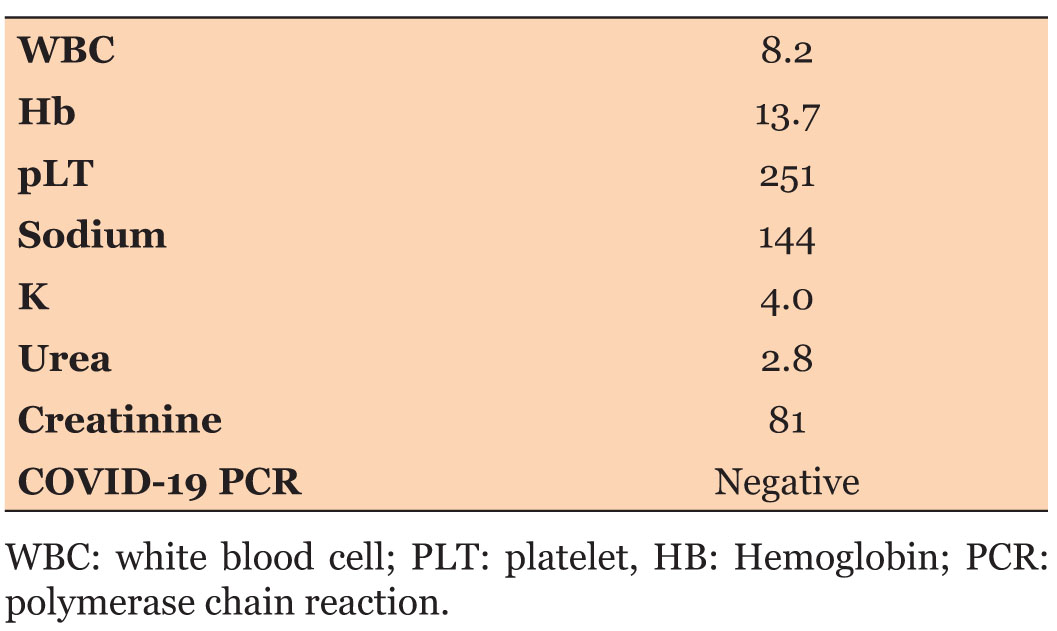
The patient reported a dollar pain for two days associated with nausea. There was no history of fever, difficulty breathing, or pleuritic chest pain. The patient was fully conscious and oriented. He was not in respiratory distress, cyanotic, or jaundiced upon examination. Because of the pain, he was clutching his abdomen with his hands. During abdominal palpation, he had localized rebound tenderness in the right iliac fossa at McBurney’s point. The patient had an occasional cough, so a COVID-19 swab was performed, which came back negative. Cell count and differential, a comprehensive metabolic panel, urine dipstick, and CRP (C-reactive protein) were within normal limits (Table 1).
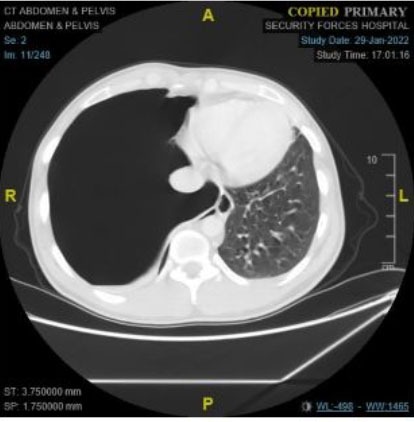
A computed tomography (CT) scan abdomen was ordered to assess for appendicitis. While reviewing the CT image, the radiologist noticed large right-sided pneumothorax. They immediately contacted the treating physician in light of this unexpected finding (Figure 1 and Figure 2). The CT report stated that there was a large pneumothorax with a leftward mediastinal shift. Two hepatic lesions (2.8×3.7 cm and 2.22 cm) with thick peripheral nodular enhancement and central hypodensity are seen in segments 2 and 4. The rest of the exam was unremarkable. There was no appendicitis.
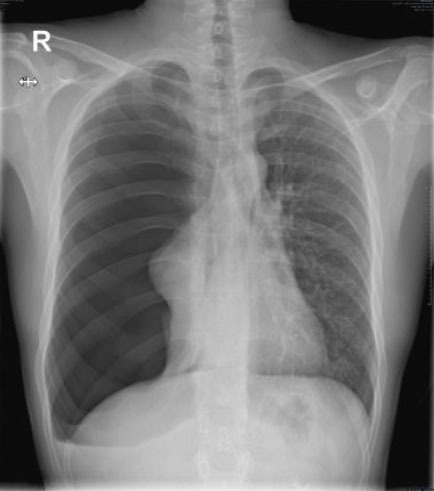
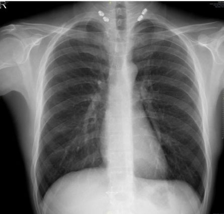
A tube thoracostomy was performed. Figure 3 shows the chest X-ray post-tube insertion. The patient was admitted to the surgical ward for monitoring and further care under thoracic surgery.
A CT chest was ordered to assess for any bullae. The CT revealed a chest tube in the right lower lobe at the superior segment, causing mild subcutaneous surgical emphysema. There was still a large right pneumothorax. There is right apical bulla measuring 0.7×2 cm. The trachea and major airways were patent, and there were small bilateral pleural effusions and bibasilar atelectasis. As a result, the patient underwent a thorascopic bullectomy and pleurodesis surgery.
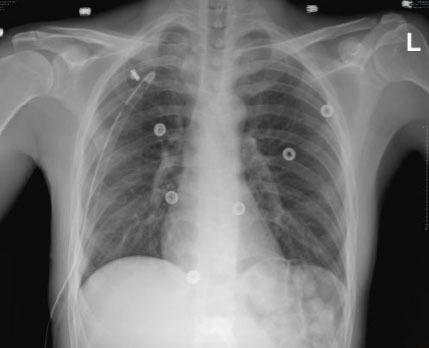
The biopsy report of the right upper lung lobe wedge resection noted lung tissue with subpleural bullae adjacent chronic inflammation with eosinophiles and mild fibrosis of the pleura. The biopsy report of the right parietal pleural excision noted fibrosis tissue with reactive mesothelial changes. The patient was discharged a few days later in a stable condition, with a follow-up outpatient clinic appointment. The last chest X-ray before discharge, as shown in Figure 4.
DISCUSSION
Pneumothorax can be fatal if it is not diagnosed and treated in a timely manner. This case could have gone unnoticed if the CT scan had not included the lower chest. Missing such diagnosis could have resulted in a serious and life-threatening situation [14].
Pneumothorax is diagnosed through a history and physical examination that typically includes pleuritic chest pain and or shortness of breath. There can be decreased air entry and hyper-resonance on percussion in the affected area. A large pneumothorax with tension physiology may result in tracheal deviation and create hemodynamic instability as a result of the increased pressure on the heart and decreased venous return [15],[16].
There are numerous examples of common diseases reported with atypical presentations in the medical literature. In one of these cases, after a few days of worsening symptoms, a patient was admitted to the hospital with community-acquired pneumonia, and the final diagnosis was confirmed to be perforated subhepatic appendicitis with abscess collection [17]. In another case, the patient reported chest pain with ischemic electrocardiogram (ECG) changes, and was later diagnosed with a perforated viscus [18].
In one study, the following risk factors for spontaneous pneumothorax were identified: male, a mean age of 24 ± 6 years, smoking, a mean body mass index (BMI) of 19.2 ± 3.8 kg/m2, and a height of 171 ± 8 cm [19]. Our patient is a smoker, 179 cm tall, 63 kg weight, and has a BMI of 19.7.
One study trying to assess if a chest X-ray should be ordered for every patient complaining of abdominal pain, ordered a chest radiograph on all patients admitted to the surgical ward (344 patients) for abdominal pain. The results revealed that 7% had a new finding and 1% had air under the diaphragm, respectively [20]. As a result, the vast majority of the X-rays that were requested were normal. This study supports the need to look for other causes of abdominal pain outside the abdomen if there are any abnormal findings on the exam or if there is no clear explanation for the patient’s symptoms.
CONCLUSION
The importance of early diagnosis of pneumothorax cannot be overstated. The majority of the time manifests as typical complaints that can be diagnosed and treated accordingly. Unfortunately, in rare instances, the presentation may be atypical.
REFERENCE
1.
Pollack CV Jr, Cantor RM, Riese VG. “Pneumothorax (Tension).” Differential Diagnosis of Cardiopulmonary Disease. Cham: Springer; 2019. p. 875–88.

2.
Huan NC, Sidhu C, Thomas R. Pneumothorax: Classification and etiology. Clin Chest Med 2021;42(4):711–27. [CrossRef]
[Pubmed]
3.
Ferguson B, Geralds J, Petrey J, Huecker M. Malpractice in emergency medicine—A review of risk and mitigation practices for the emergency medicine provider. J Emerg Med 2018;55(5):659–65. [CrossRef]
[Pubmed]
4.
Osterwalder I, Özkan M, Malinovska A, Nickel CH, Bingisser R. Acute abdominal pain: Missed diagnoses, extra-abdominal conditions, and outcomes. J Clin Med 2020;9(4):899. [CrossRef]
[Pubmed]
5.
Chen J(Steven), Kandle PF, Murray I, Fitzgerald LA, Sehdev JS. Physiology, Pain. 2021 Jul 26. In: StatPearls. Treasure Island (FL): StatPearls Publishing; 2022.
[Pubmed]
6.
Brikman S, Levi O, Dori G. Rare clinical manifestation of community-acquired pneumonia. BMJ Case Rep 2018;11(1):e225589. [CrossRef]
[Pubmed]
7.
Mumtaz H, Mustafa K, Safdar K, Meer F, Fatima T, Shafiq MA. Invisible or spontaneous pneumothorax: An unusual presentation. Biomed J Sci & Tech Res 2020;30(5):23786–8. [CrossRef]
8.
Suwanwongse K, Shabarek N. Pseudo-appendicitis in an adolescent with COVID-19. Cureus 2020;12(7):e9394. [CrossRef]
[Pubmed]
9.
Soaid MS, Ahmad N. Spontaneous pneumothorax mimicking perforated viscus in Covid-19 patient. BJOSS 2021;1(1):8–13.
10.
Han Y, Gong Y. Pulmonary embolism with abdominal pain as the chief complaint: A case report and literature review. Medicine (Baltimore) 2019;98(44):e17791. [CrossRef]
[Pubmed]
11.
Sukumar SM. An atypical tension pneumothorax. Clin Med (Lond) 2020;20(Suppl 2):s52. [CrossRef]
[Pubmed]
12.
Sanvictores T, Tadi P. Neuroanatomy, Autonomic Nervous System Visceral Afferent Fibers and Pain. 2021 Oct 9. In: StatPearls. Treasure Island (FL): StatPearls Publishing; 2022.
[Pubmed]
13.
Morrone D, Morrone V. Acute pulmonary embolism: Focus on the clinical picture. Korean Circ J 2018;48(5):365–81. [CrossRef]
[Pubmed]
14.
Wang XH, Duan J, Han X, et al. High incidence and mortality of pneumothorax in critically ill patients with COVID-19. Heart Lung 2021;50(1):37–43. [CrossRef]
[Pubmed]
15.
Jalota Sahota R, Sayad E. Tension Pneumothorax. 2022 May 12. In: StatPearls. Treasure Island (FL): StatPearls Publishing; 2022.
[Pubmed]
16.
Zietlow J, Hernandez M, Bestland A, et al. Decompression of tension pneumothorax in a trauma patient – First use of a novel decompression colorimetric capnography device in human patient. Gen Thorac Cardiovasc Surg 2021;69(2):391–3. [CrossRef]
[Pubmed]
17.
Pires JR, Moreira MJ, Martins M, Neves C. Unusual pneumonia mimic. Eur J Case Rep Intern Med 2019;6(7):001181. [CrossRef]
[Pubmed]
18.
Secchi MF, Torre C, Dui G, Virdis F, Podda M. The close relationship between large bowel and heart: When a colonic perforation mimics an acute myocardial infarction. Case Rep Surg 2018;2018:8020197. [CrossRef]
[Pubmed]
19.
Aljehani YM, Almajid FM, Niaz RC, Elghoneimy YF. Management of primary spontaneous pneumothorax: A single-center experience. Saudi J Med Med Sci 2018;6(2):100–3. [CrossRef]
[Pubmed]
20.
Alazzawi S, De Rover WS, Morris-Stiff G, Lewis MH. Erect chest radiography in the setting of the acute abdomen: Essential tool or an unnecessary waste of resources? Ann R Coll Surg Engl 2010;92(8):697–9. [CrossRef]
[Pubmed]
SUPPORTING INFORMATION
Acknowledgement
The authors thank Dr. Gary Vilke for editing the manuscript.
Author ContributionsYahia Yaseen Akeely - Conception of the work, Design of the work, Acquisition of data, Analysis of data, Drafting the work, Revising the work critically for important intellectual content, Final approval of the version to be published, Agree to be accountable for all aspects of the work in ensuring that questions related to the accuracy or integrity of any part of the work are appropriately investigated and resolved.
Abdelwahed Syyar Alenezy - Conception of the work, Design of the work, Acquisition of data, Analysis of data, Drafting the work, Revising the work critically for important intellectual content, Final approval of the version to be published, Agree to be accountable for all aspects of the work in ensuring that questions related to the accuracy or integrity of any part of the work are appropriately investigated and resolved.
Sultan Mohammed Al Marzouqi - Conception of the work, Design of the work, Acquisition of data, Analysis of data, Drafting the work, Revising the work critically for important intellectual content, Final approval of the version to be published, Agree to be accountable for all aspects of the work in ensuring that questions related to the accuracy or integrity of any part of the work are appropriately investigated and resolved.
Nader Bokhari - Conception of the work, Design of the work, Acquisition of data, Analysis of data, Drafting the work, Revising the work critically for important intellectual content, Final approval of the version to be published, Agree to be accountable for all aspects of the work in ensuring that questions related to the accuracy or integrity of any part of the work are appropriately investigated and resolved.
Mohammad Yousef - Conception of the work, Design of the work, Acquisition of data, Analysis of data, Drafting the work, Revising the work critically for important intellectual content, Final approval of the version to be published, Agree to be accountable for all aspects of the work in ensuring that questions related to the accuracy or integrity of any part of the work are appropriately investigated and resolved.
Guarantor of SubmissionThe corresponding author is the guarantor of submission.
Source of SupportNone
Consent StatementWritten informed consent was obtained from the patient for publication of this article.
Data AvailabilityAll relevant data are within the paper and its Supporting Information files.
Conflict of InterestAuthors declare no conflict of interest.
Copyright© 2022 Yahia Yaseen Akeely et al. This article is distributed under the terms of Creative Commons Attribution License which permits unrestricted use, distribution and reproduction in any medium provided the original author(s) and original publisher are properly credited. Please see the copyright policy on the journal website for more information.


