 |
|
Case Report
| ||||||
| Mesenchymal cystic hamartoma of the lung associated with diffuse alveolar damage | ||||||
| Luvo Gaxa1, Bafana Hlatshwayo2, Aletta Lefentse Motene3 | ||||||
|
1MBChB, MMed (Rad D), Pediatric Radiology Fellow, Red Cross war Memorial Children’s Hospital Coromplex, Polokwane, Capetown, South Africa
2Dip Rad Diag, MBChB, MMed (Rad D), Senior Specialist, Diagnostic Radiology and Imaging, Polokwane-Mankweng Hospital Complex, Polokwane, Limpopo, South Africa 3MBchB, FC Ped, Fellow of Pediatric Pulmonology, Senior Specialist, Pediatrics, Polokwane-Mankweng Hospital Complex, Polokwane, Limpopo, South Africa | ||||||
| ||||||
|
[HTML Abstract]
[PDF Full Text]
[Print This Article]
[Similar article in Pumed] [Similar article in Google Scholar] |


| How to cite this article |
| Gaxa L, Hlatshwayo B, Motene AL. Mesenchymal cystic hamartoma of the lung associated with diffuse alveolar damage. Case Rep Int 2017;6:27–30. |
|
ABSTRACT
|
|
Introduction:
Mesenchymal cystic hamartoma (MCH) of the lung is a clinicopathological and a rare entity with typical pathological features. Mesenchymal cystic hamartoma is characterized by cystic pulmonary lesions that tend to increase in size slowly. The pulmonary hamartomas usually are asymptomatic more especially when they are small in size. The treatment is conservative in asymptomatic patients. The ultimate treatment is surgical for the large and symptomatic lung mesenchymal hamartomas. Case Report: A six-month-old male neonate was referred from a regional hospital with a history of progressive shortness of breath and fever. The chest X-ray was performed and it showed a thin walled left lung cystic mass with mediastinal deviation to the right side. Computed tomography scan followed the chest X-ray and it confirmed large and smaller cysts with thin walls. Biopsy was performed and it confirmed the diagnosis of mesenchymal cystic hamartoma of the lung. Thoracotomy and the excision of the left lung cysts were done and later the neonate was discharged from the hospital without complications. Conclusion: It is of utmost importance to consider a cystic mesenchymal lung hamartoma in our differential diagnosis when dealing with cystic lung masses in children. | |
|
Keywords:
Clinicopathological, Mesenchymal cystic lung hamartoma, Treatment
| |
|
INTRODUCTION
| ||||||
|
Mesenchymal cystic lung hamartoma is a rare benign cystic mass of the lungs with no specific clinical or radiological signs. Pathologically this condition typically presents as the cysts that have a tendency to grow slowly over time. There is a possibility of misdiagnosing a mesenchymal cystic hamartoma as a cystic pleuropulmonary blastoma or as a congenital pulmonary adenoid malformation. It is important to note that the mesenchymal hamartomas are not only limited to the lungs but may also rarely occur in the chest wall and the liver. | ||||||
|
CASE REPORT
| ||||||
|
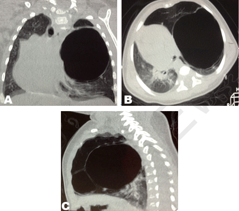
A six-month-old male neonate was referred from a regional hospital with a history of progressive shortness of breath and fever. Previously, a two-month history of cough and the patient was treated as having pneumonia in the regional hospital. There was no history of loss of appetite. The mother of the patient is a 25-year-old para1 confirmed Human immuno-deficiency negative (HIV-) who had an uneventful pregnancy. The baby was born at term with a normal vaginal delivery with no complications at birth. The birth weight 2900g, apgar sore 10/10, head circumference 34 cm, birth height 46 cm. The neonatal period was uneventful. The immunization status is up to date. The patient has a normal growth and development. On examination the patient had moderate tachypnea. Oxygen 0.5 L/min was given via the nasal prongs .The air-entry was decreased with hyper-resonance on the left lung. The right lung was within normal limits. The cardiac apex beat was palpable on the right sided presumable due to mediastinal deviation to the right side. The laboratory examinations showed a normal full blood count, normal urea and electrolytes. The erythrocyte sedimentation rate 20 mm/hr, C-reactive protein less than 1 mg/dl. The chest X-ray was performed and it demonstrated multiple round to oval shaped cystic lesions in the left lung with associated mediastinal deviation to the right side (Figure 1). Computed tomography (CT) scan of the chest was subsequently performed to further characterize the chest radiograph findings. Computed tomography scan confirmed multiple left lung cystic masses and congenital pulmonary adenomatoid malformation was the working diagnosis following CT scan. The larger lesion measured 9x7.3 cm and the smaller lesion measured 3.2x3 cm on axial view on widest diameter (Figure 2). Biopsy was performed and the histopathology report was as follows: Microscopy: Section taken from both specimens I and II show lung tissue with features of mesenchymal cystic hamartoma associated with diffuse alveolar damage. There were multifocal nodular endo-bronchial or bronchiolar growths covered by non-neoplastic epithelium. The tumor consists of lobules of cartilage, fat, fibromyxoid tissue, and sometimes smooth muscle that are separated by clefts lined by non-neoplastic respiratory epithelium. The distal air-spaces show features of diffuse alveolar damage. The air spaces are obliterated by fibrinous exudates with a few desquamated pneumocytes present. The final diagnosis is mesenchymal cystic lung hamartoma associated with diffuse alveolar damage. Thoracotomy and the excision of the left lung cysts were done and later the patient was discharged from the hospital without complications. On recent follow-up the baby is growing up well with no clinical abnormalities (Figure 3) | ||||||
| ||||||
| ||||||
| ||||||
|
DISCUSSION | ||||||
|
Mesenchymal cystic hamartoma of the lung is a clinicopathological and a rare entity with typical pathological features that is characterized by cystic pulmonary lesions that tend to increase in size slowly [1]. By the year 2012 only less than 15 cases were reported since year 1986 in English literature but the exact incidence of this extremely rare disease remains unknown [1][2]. The very first reports of mesenchymal cystic hamartoma were recorded by the year 1986 by Mark [2]. The pulmonary hamartomas usually are asymptomatic more especially when they are small in size and the treatment is conservative if asymptomatic but the ultimate treatment is surgical for the large and symptomatic lung mesenchymal hamartomas [3]. The female to male ratio is 11:4 and the reported age of occurrence ranges between 1.5 and 53 years of age [4]. Although not specific the clinical presentation may entail but not limited to pleuritic chest pain, mild to moderate dyspnea, hemoptysis and hemothorax [4]. The lack of specific clinical symptoms of mesenchymal cystic hamartoma warrants a need for the attending physician to know the following differential diagnosis and their features [4]:
It is also important to have additional differential diagnosis when dealing with MCH such as bronchopulmonary sequestration, bronchiectasis and endometrial stromal sarcoma metastasis [4]. Hemoptysis is a complication which may be lethal and results from hemorrhage from the systemic arteries into the cysts and other complications of mesenchymal cystic hamartoma are the pneumothorax and the hemothorax which occur as a result of rupture of sub-pleural cyst [4]. It is important to note that the mesenchymal hamartomas are not only limited to the lungs but may also occur in the chest wall and the liver [5]. The chest wall hamartoma leads to a severely deformed thorax and commonly involves the pleural ribs and vertebrae [6]. In mesenchymal cystic hamartomas (MCH), nodules occur first and when the nodules increase in size they become cystic [7]. Mesenchymal cystic hamartomas disrupts the normal lung architecture and the spectrum of presentation is broad and may range from [7]:
(a) nodular disease that is not progressive and One case report showed pleural and diaphragmatic invasion by MCH seen on magnetic resonance imaging scan and Positron emission Computed tomography (PET CT) scan [8]. | ||||||
|
CONCLUSION
| ||||||
|
The ultimate diagnosis of mesenchymal cystic hamartoma is though biopsy. The main management of these lesions is surgical if they are symptomatic. | ||||||
|
REFERENCES
| ||||||
| ||||||


|
[HTML Abstract]
[PDF Full Text]
|
|
Acknowledgements
We are thankful to our radiographers Molopa Moyahabo Tink, Makuka Emilly Mogashoa, Julia Madzorera and Portia Macheke for retrieving and preparing images for this case study. |
|
Author Contributions
Luvo Gaxa – Substantial contributions to conception and design, Acquisition of data, Analysis and interpretation of data, Drafting the article, Revising it critically for important intellectual content, Final approval of the version to be published Bafana Elliot Hlatshwayo – Substantial contributions to conception and design, Revising it critically for important intellectual content, Final approval of the version to be published Aletta Lefentse Motene – Analysis and interpretation of data, Revising it critically for important intellectual content, Final approval of the version to be published |
|
Guarantor of submission
The corresponding author is the guarantor of submission. |
|
Source of support
None |
|
Conflict of interest
Authors declare no conflict of interest. |
|
Copyright
© 2017 Luvo Gaxa et al. This article is distributed under the terms of Creative Commons Attribution License which permits unrestricted use, distribution and reproduction in any medium provided the original author(s) and original publisher are properly credited. Please see the copyright policy on the journal website for more information. |
|
|