|
|
|
Case Report
|
| |
|
A rare case of fulminant primary Epstein–Barr virus infection in a young adult
|
|
Jason Tan1,
Chaminda Basnayake2
|
1Senior Registrar, Division of Medicine, The Queen Elizabeth Hospital, Adelaide, South Australia, Jason.
2Staff Consultant, Division of Medicine, The Queen Elizabeth Hospital, Adelaide, South Australia.
|
Article ID: 100017CRINTJT2015
doi:10.5348/crint-2015-17-CR-12
|
Address correspondence to:
Jason Jun'an Tan
The Queen Elizabeth Hospital. 28 Woodville Road
Woodville South
South Australia
Australia 5011
Phone: +6182226000
Fax: +6182226126
|
Access full text article on other devices
 |
|
Access PDF of article on other devices
 |
|
|
[HTML Abstract]
[PDF Full Text]
[Print This Article]
[Similar article in Pumed]
[Similar article in Google Scholar]
|
|
How to cite this article:
|
|
Tan J, Basnayake C. A rare case of fulminant primary Epstein–Barr virus infection in a young adult. Case Rep Int 2015;4:47–51.
|
|
Introduction:
Primary Epstein–Barr virus (EBV) infection usually manifests in adolescents and young adults as infectious mononucleosis which classically presents as a syndrome of fever, pharyngitis, lymphadenopathy, fatigue and atypical lymphocytosis. The vast majority of patients have relatively mild disease. However, it may rarely present as a fulminant infection with severe multi-organ manifestations.
Case Report:
We report a rare case of a young woman admitted under our care in hospital with severe primary EBV infection. A 20-year-old female with no significant past medical history presented with a three-week history of worsening sore throat, ear pain, non-productive cough, fever, nausea, generalized myalgia and lethargy. Serology confirmed that she had acute EBV infection. During her admission, she developed complications of fulminant hepatic failure, coagulopathy, retropharyngeal abscess, ulcerative glossitis and pulmonary consolidation resulting in hemoptysis and sepsis which required intensive care unit management. With aggressive antibiotic and supportive therapy, she made a full recovery.
Conclusion:
This is the first case report documenting the concurrent presence of severe multi-system involvement in primary EBV infection including hepatic, haematological, upper and lower respiratory tract. Despite the benign course of disease in the vast majority of patients with primary EBV infection, this case highlights the need for clinicians to be aware of the possibility of fulminant disease with severe multi-organ involvement with potential life-threatening consequences. Clinicians treating patients with primary EBV infection should remain vigilant in monitoring and treating severe complications.
|
Keywords:
Coagulopathy, Epstein-Barr virus, Haemoptysis, Infection, Liver failure, Retropharyngeal abscess
|
Introduction
|
|
Epstein–Barr virus (EBV) is a widely disseminated gamma herpesvirus which was first identified in 1964 [1]. It is spread by intimate contact between susceptible persons and EBV shedders, primarily via saliva [2]. Primary EBV infection during childhood is often subclinical, however from adolescence through to early adulthood, the incidence of symptomatic infection increases and is most commonly associated with infectious mononucleosis (IM). The classic features of IM include fever, pharyngitis, lymphadenopathy, fatigue and atypical lymphocytosis [3]. The majority of patients with IM recover from their initial symptoms (e.g., fever, sore throat, lymphadenopathy) by 1 month but a minority have persistent fatigue which may last for more than 6 months [4]. The vast majority of patients have relatively mild disease however rarely, acute serious complications may occur. These include hematologic or neurological disease, hemophagocytic lymphohistiocytosis, splenic rupture and upper airway obstruction from lymphoid hyperplasia and mucosal oedema [5]. We present a rare case of a 20-year-old female with primary EBV infection complicated by fulminant hepatic failure, coagulopathy, retropharyngeal abscess, ulcerative glossitis and pulmonary consolidation.
|
Case Report
|
|
A 20-year-old unemployed Caucasian female from rural New South Wales was admitted to hospital with a three-week history of worsening sore throat affecting oral intake, left ear pain, non-productive cough, fever, nausea, generalized body aches and lethargy. Her past medical history included migraine, attention-deficit hyperactivity disorder and tonsillectomy. She was not on any regular medications and had no significant smoking, alcohol or illicit drug use. She had previously presented to the emergency department of a different peripheral hospital eight days prior with similar symptoms and was diagnosed with Epstein-Barr virus (EBV) infection with a positive serum monospot test.
Initial physical examination revealed a low-grade fever with swollen tender left submandibular and cervical lymph nodes. Serology confirmed acute EBV infection with both EBV IgM and IgG present. Blood picture showed lymphocytosis (5.31x10⁹/L) with atypical lymphocytes seen on blood film examination. Liver function tests were acutely deranged with a predominantly hepatocellular pattern with impaired synthetic function: bilirubin 2.5 mg/dL, GGT 173 U/L, ALP 184 U/L, ALT 933 U/L, AST 1478 U/L, albumin 34 g/L, PT 22.9s, INR 2.0. Paracetamol level (120 µmol/L) was within the therapeutic range and urine beta-hCG was negative. Testing for viral and autoimmune hepatitis was negative. Initial chest radiograph and urine culture were unremarkable.
The patient was initially treated with supportive measures including analgesia, antiemetics, intravenous fluids and vitamin K. Monitoring of liver function tests showed further worsening. On day-3 of admission: bilirubin 3.9 mg/dL, GGT 194 U/L, ALP 181 U/L, ALT 3950 U/L, AST 5060 U/L, albumin 24 g/L, PT 32.6s, INR 3.1. On day-4 of admission, her fever worsened to 38.7°C and she had 400 ml of hemoptysis. Prothrombinex and vitamin K were administered. Emergency upper gastrointestinal endoscopy was unremarkable apart from a blood clot at the epiglottis and clotted blood in stomach with no source of bleeding identified.
Subsequently, she was intubated and transferred to the intensive care unit. Bronchoscopy was normal. Microlaryngoscopy revealed necrosis, sloughing and ulceration of left side of tongue base, vallecular, tonsillolingual sulcus, aryepiglottic fold up to arytenoid. In addition, there was lateral wall pyriform inflammation and sloughing. Biopsies were consistent with ulcerative glossitis with no evidence of malignancy or granulomata. In-situ hybridization revealed occasional EBV positive cells. Swab culture was negative for bacteria.
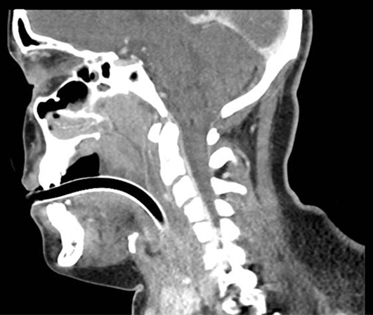
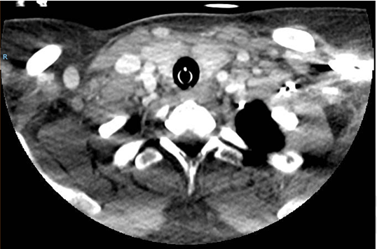
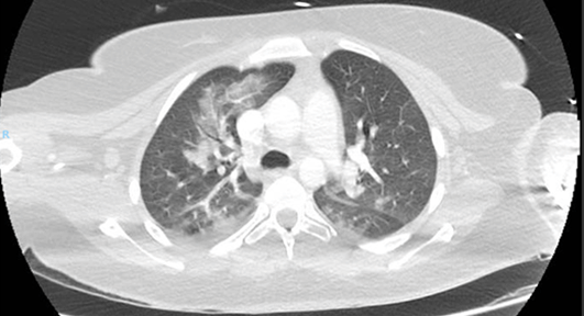
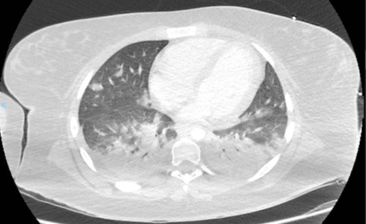
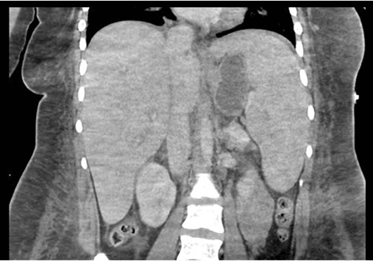
Computed tomography scan of the neck, chest and abdomen (Figure 1) (Figure 2) (Figure 3)
(Figure 4) (Figure 5) revealed multiple findings: fluid filled pharynx with a fluid collection at the retropharyngeal region measuring up to 6 mm in thickness, from the level of the C1 to C4/5 level; widespread lymphadenopathy involving both anterior and posterior cervical chain; bi-basal consolidation posteriorly, mixed ground-glass opacity and consolidative changes at the perihilar region and small right basal effusion; extensive abdominal soft tissue subcutaneous oedema; hepatosplenomegaly (20 cm/20 cm).
The patient was transfused 1 unit of red blood cells and commenced on intravenous piperacillin/tazobactam which was subsequently changed to intravenous metronidazole, clindamycin and benzylpenicillin for wider microbial cover including anaerobes. Video laryngoscope performed on day-4 of ICU admission showed ongoing purulent discharge into oropharynx with improvement in edema of epiglottis, aryepiglottic fold and tongue base. Given the presence of fistula draining the retropharyngeal abscess, surgical drainage was not performed. Nasogastric tube was inserted during that time and enteral feeds were commenced. She was extubated on day-7 of ICU admission after bedside laryngoscopy confirmed further decrease in edema and transferred to a general ward the following day.
Given her clinical recovery, antibiotics were ceased after a 10-day course. Oral diet was commenced and her nasogastric tube was removed. She received input from Allied Health teams including physiotherapy and speech pathology. Her fever and lymphadenopathy resolved. Liver function continued to improve. At time of discharge, her liver function tests were as follows: bilirubin 1.2 mg/dL, GGT 134U/L, ALP 65 U/L, ALT 92 U/L, AST 49 U/L, albumin 26 g/L, PT 35s, INR 1.1. The patient was discharged home on day-14 of admission.
|
|
|
|
|
|
|
|
|
|
|
|
|
|
Discussion
|
|
We report a rare case of a 20-year-old female with fulminant EBV infection complicated by severe hepatic failure, coagulopathy, retropharyngeal abscess, ulcerative glossitis and pulmonary consolidation that was successfully managed. To our knowledge, this is the first case report documenting the concurrent presence of severe multi-system involvement in primary EBV infection: hepatic, hematological, upper and lower respiratory tract.
Although mild hepatitis is common feature in IM with minor elevations in serum transaminases, severe liver injury is extremely rare. This is usually related to congenital or acquired immunodeficiency syndromes, including HIV, complement deficiency, X-linked lymphoproliferative disease or cancer chemotherapy [6]. Histopathological findings include lymphocytic hepatic infiltration and Kupffer cell proliferation resulting in mild intrahepatic cholestasis but maintenance of the lobular architecture and without necrosis [7]
. There was no prior history of significant infections to suggest that congenital or acquired immunodeficiency was present in our patient.
The most common hematologic finding in IM is lymphocytosis. Atypical lymphocytes may be detected. Unusual hematologic manifestations include hemolytic anemia, thrombocytopenia, aplastic anemia, thrombotic thrombocytopenic purpura/hemolytic-uremic syndrome, and disseminated intravascular coagulation [8]. Our patient developed a coagulopathy with prolonged PT mostly as a result of her deranged hepatic synthetic function. Other potential contributors include dietary factors and use of broad spectrum antibiotics.
Upper airway complications e.g., peritonsillar abscess have been reported as a rare complication of IM. It has been estimated that peritonsillar abscess account for 1% of patients with IM who are admitted to hospital [9]. Early case reports have reported a possible association with corticosteroid use, but this link is controversial [10]. Our patient did not have any corticosteroid use. Intrathoracic involvement in IM, especially pulmonary consolidation, is uncommon. The most common pulmonary radiological finding are mediastinal lymphadenopathy and rarely, interstitial pulmonary infiltrate [11]. Histopathological features include mononuclear inflammatory cells present along bronchovascular bundles and interlobular septa in interstitial pulmonary infiltrates [12].
Our patient developed acute upper airway bleeding requiring blood transfusion and ICU admission. Bleeding has not been previously reported as a complication of IM until a recent case report which presented an 18-year-old male with IM complicated by spontaneous gastric and esophageal bleeding with necrosis and subsequent perforation [13]. Factors for the upper airway bleeding in our patient included coagulopathy from liver failure and ulcerative glossitis.
Other than antibiotic therapy, our patient received mainly supportive treatment with close monitoring. We did not use corticosteroids or antiviral medications are there is no clear evidence for their efficacy. Over the 14 days of admission, her IM complications had largely resolved without any serious sequelae.
|
Conclusion
|
|
We have reported a case of fulminant primary EBV infection with potentially life-threatening multi-organ complications in a healthy young adult. Despite the benign course of disease in the vast majority of patients with infectious mononucleosis, this case highlights the need for clinicians to be aware of the possibility of fulminant disease with severe multi-organ involvement. Clinicians treating patients with primary Epstein–Barr Virus (EBV) infection should remain vigilant in monitoring the development of severe complications.
|
References
|
-
Jenson HB. Ebstein-Barr virus. Ped in Rev 2011;32(9):275–384.

-
Balfour HH Jr, Holman CJ, Hokanson KM, et al. A prospective clinical study of Epstein-Barr virus and host interactions during acute infectious mononucleosis. J Infect Dis 2005 Nov 1;192(9):1505–12.
[Pubmed]
-
Luzuriaga K, Sullivan JL. Infectious mononucleosis. N Engl J Med 2010 May 27;362(21):1993–2000.
[CrossRef]
[Pubmed]
-
Rea TD, Russo JE, Katon W, Ashley RL, Buchwald DS. Prospective study of the natural history of infectious mononucleosis caused by Epstein-Barr virus. J Am Board Fam Pract 2001 Jul-Aug;14(4):234–42.
[Pubmed]
-
Evans A, Niederman J. Epstein-Barr virus. In: Evans A (Editor). Viral Infections of Human Epidemiology and Control. New York: Plenum Publishing; 1989. p. 265.
-
Feranchak AP, Tyson RW, Narkewicz MR, Karrer FM, Sokol RJ. Fulminant Epstein-Barr viral hepatitis: orthotopic liver transplantation and review of the literature. Liver Transpl Surg 1998 Nov;4(6):469–76.
[CrossRef]
[Pubmed]
-
Jenson HB. Acute complications of Epstein-Barr virus infectious mononucleosis. Curr Opin Pediatr 2000 Jun;12(3):263–8.
[CrossRef]
[Pubmed]
-
Auwaerter PG. Infectious mononucleosis in middle age. JAMA 1999 Feb 3;281(5):454–9.
[CrossRef]
[Pubmed]
-
Johnsen T. Infectious mononucleosis and peritonsillar abscess. J Laryngol Otol 1981 Aug;95(8):873–6.
[CrossRef]
[Pubmed]
-
Hanna BC, McMullan R, Hall SJ. Corticosteroids and peritonsillar abscess formation in infectious mononucleosis. J Laryngol Otol 2004 Jun;118(6):459–61.
[CrossRef]
[Pubmed]
-
Miyake H, Matsumoto A, Komatsu E, et al. Infectious mononucleosis with pulmonary consolidation. J Thorac Imaging 1996 Spring;11(2):158–60.
[CrossRef]
[Pubmed]
-
Myers JL, Peiper SC, Katzenstein AL. Pulmonary involvement in infectious mononucleosis: histopathologic features and detection of Epstein-Barr virus-related DNA sequences. Mod Pathol 1989 Sep;2(5):444–8.
[Pubmed]
-
Busch D, Hilswicht S, Schöb DS, et al. Fulminant Epstein-Barr virus - infectious mononucleosis in an adult with liver failure, splenic rupture, and spontaneous esophageal bleeding with ensuing esophageal necrosis: a case report. J Med Case Rep 2014 Feb 5;8:35.
[CrossRef]
[Pubmed]
|
[HTML Abstract]
[PDF Full Text]
|
|
Author Contributions
Jason Tan – Conception and design, Acquisition of data, Analysis and interpretation of data, Drafting the article, Critical revision of the article, Final approval of the version to be published
Chaminda Basnayake – Drafting the article, Critical revision of the article, Final approval of the version to be published
|
Guarantor of submission
The corresponding author is the guarantor of submission.
|
Source of support
None
|
Conflict of interest
Authors declare no conflict of interest.
|
Copyright
©
2015 Jason Tan et al. This article is distributed under the terms of Creative Commons Attribution License which permits unrestricted use, distribution and reproduction in any medium provided the original author(s) and original publisher are properly credited. Please see the copyright policy on the journal website for more information.
|
|
|