| Table of Contents | |
|
Case Report
| ||||||
| Vallecular cyst | ||||||
| Luvo Gaxa1, Bafana Elliot Hlatshwayo2, Magaroleng Hendry Modishi3 | ||||||
|
1MBChB, Senior Registrar, Diagnostic Radiology and Imaging, Polokwane-Mankweng Hospital Complex, Polokwane, Limpopo, South Africa.
2Dip Rad Diag, MBChB, MMed Rad D, Senior Specialist, Diagnostic Radiology and Imaging, Polokwane- Mankweng Hospital Complex, Polokwane, Limpopo, South Africa. 3MBChB, MMed Rad D, FC Rad D, Principal Specialist, Diagnostic Radiology and Imaging, Polokwane-Mankweng Hospital Complex, Polokwane, Limpopo, South Africa. | ||||||
| ||||||
|
[HTML Abstract]
[PDF Full Text]
[Print This Article]
[Similar article in Pumed] [Similar article in Google Scholar] |
| How to cite this article: |
| Gaxa L, Hlatshwayo BE, Modishi MH. Vallecular cyst. Case Rep Int 2015;4:6–10. |
|
Abstract
|
|
Introduction:
A vallecular cyst is a rare and a benign entity which is readily diagnosed in newborns and in early childhood and is associated with a high morbidity and mortality. The symptoms of a vallecular cyst include but not limited to failure to thrive, voice changes, feeding difficulties, stridor and shortness of breath while in adults the symptoms are commonly mild and the vallecular cyst in the latter tends to be diagnosed as an incidental finding. As early as the prenatal period, vallecular cysts can be diagnosed by using either ultrasonography or magnetic resonance imaging (MRI) scan, MRI scan proves superior and is highly recommended to diagnose and to obtain thorough information regarding the relationship of the cyst to the surrounding anatomical structures and that influences the patient's treatment greatly.
Case Report: We report a case of a seven-year-old boy referred from the peripheral hospital presented with snoring kind of breathing since six months ago. The patient had a difficulty in breathing when sleeping. The patient was able to take meals without any difficulty. No past medical history reported. On examination the oro-pharyngeal mass was noted. Blood pressure 105/57 mmHg, pulse 105 /minute, temperature 36.5°C, weight 13 kg, height 114 cm. The patient had normal developmental milestones. On laryngoscope, a cystic mass in the vallecular region measuring 2.4×2.7 cm and the mass was attached to the base of the tongue, pharyngeal wall and the lingual surface of the epiglottis. The diagnosis on left side vallecular cyst was made on clinical and radiological findings. The cyst was treated by marsupialization and the specimen was sent to the laboratory which confirmed a diagnosis of a vallecular cyst. There was no recurrence of a cyst on our patient's follow-up. Conclusion: A vallecular cyst shows some typical imaging findings on computed tomography scan and on magnetic resonance imaging scan and a multi-disciplinary approach helps to secure the diagnosis. | |
|
Keywords:
Breathing difficulty, Snoring, Marsupialization, Ultrasound, Vallecular cyst
| |
|
Introduction
| ||||||
|
Although a vallecular cyst is rare, it is a well known cause of airway-obstruction that occasionally leads to stridor, cough, dysphagia, dysphonia, foreign body sensation and death, and is readily diagnosed but not only limited to newborns and infants [1] [2] [3]. As early as the prenatal period, vallecular cysts can be diagnosed by using either ultrasonography or magnetic resonance imaging (MRI) scan, MRI scan proves superior and is highly recommended to diagnose and to obtain thorough information regarding the relationship of the cyst to the surrounding anatomical structures and that influences the patient's treatment greatly. It is difficult to estimate the exact incidence of vallecular cysts, however, the reported incidence on laryngoscopy ranges between 1 in 1,250 to 1 in 4,200 people and 10% of the population [4] [5]. Vallecular cyst displaces the epiglottis infero-posteriorly obstructing the supraglottic space and the airway obstruction also results from the mass effect the cyst exerts to the hypo-pharynx [3]. | ||||||
|
Case Report
| ||||||
|
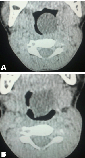
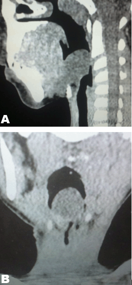
A seven-year-old boy referred from the peripheral hospital presented with snoring kind of breathing since six months ago. The patient had a difficulty in breathing when sleeping. The patient was able to take meals without any difficulty. No past medical history reported. On laryngoscopy examination an egg-shaped pink mass was seen in the oropharynx with extension towards the oral cavity. Nasal cavities and the turbinates were within normal limits. Ultrasound was then requested and it revealed a cystic neck mass. Computed tomography (CT) scan of the neck was performed following an ultrasound report. Computed tomography scan showed a left vallecular cyst which was not separable from the base of the tongue. The cyst measured 7x6 cm on axial view on its widest diameter (Figure 1) and (Figure 2). The cyst was surgically removed two weeks later and there were no postoperative complications reported and the patient was later discharged from the hospital without any symptoms (Figure 3). The surgical steps that were followed in our case are as follows:
The histopathology report was as follows: Microscopy: The section showed fragments of a cyst wall lined by a squamous cell epithelium. The wall shows a prominent lymphocytic infiltrate which led to a histopathology report of a vallecular cyst. | ||||||
| ||||||
| ||||||
| ||||||
|
Discussion
| ||||||
|
Although a vallecular cyst is rare, it is a well known cause of airway-obstruction that occasionally leads to stridor, cough, dysphagia, dysphonia, foreign body sensation and death and is readily diagnosed but not only limited to newborns and infants [1] [2] [3]. It is difficult to estimate the exact incidence of vallecular cysts, however, the reported incidence on laryngoscopy ranges between 1 in 1,250 to 1 in 4,200 people and 10% of the population [4] [5]. Vallecular cyst displaces the epiglottis infero-posteriorly obstructing the supraglottic space and the airway obstruction also results from the mass effect the cyst exerts to the hypopharynx [3]. In literature, different names for the vallecular cyst are recognised and these entail epiglottic cyst, ductal cyst, base of the tongue cyst as well as mucous retention cyst and are commonly seen if the maxillary sinus floor [1] [5]. Knowing the differential diagnosis of the vallecular cyst is of utmost importance as the management of these lesions differs completely from that of the vallecular cyst and these entities include [6]: Hemangiomas, lymphangiomas, teratomas, dermoid cyst, lingual thyroid and the internal thyroglossal duct cyst. Vallecular cysts simple occur as a result of mucous gland obstruction within the mucosal lining and that leads to cyst formation and continued secretion is responsible for the increasing size of the cyst [4] [7]. It is also hypothesized that lymphatic malformation, angiomatous (embryological malformation) as some of the potential causes of a vallecular cyst [1]. As early as the prenatal period, vallecular cysts can be diagnosed by using either ultrasonography or magnetic resonance imaging (MRI) scan and MRI scan proves superior and is highly recommended to diagnose and to obtain thorough information regarding the relationship of the cyst to the surrounding anatomical structures and that influences the patient's treatment greatly [1]. Ultrasound plays a role in diagnosing the vallecular cyst which presents as a non-vascular homogeneous hypoechoic mass with imperceptible wall and the mass may be visualized below as well as behind the tongue [8]. In cases of a complicated vallecular cyst, ultrasound may show septations or echoes within the cyst and the cyst wall may be thick [8]. On computed tomography scan (CT) simple vallecular cyst typically presents as a thin-walled hypodense lesion that does not enhance post contrast [9]. Hemorrhage and proteinaceous fluid are some of the recognized complications of the vallecular cyst on CT scan and in such cases the cyst may present as a soft tissue mass [9]. Magnetic resonance imaging (MRI) sequences used in diagnosing a vallecular cyst include a T1-weighted, T2- weighted sequences with or without gadolinium [10]. A simple vallecular cyst demonstrates homogeneous hypo-intense signal intensity on T1-weighted images and hyper-intense signal intensity on T2-weighted images and does not enhance post gadolinium except in complicated cases whereby the T1-weighted images show a hyper-intense signal intensity of a cyst [9]. There is a likely-hood of a vallecular cyst recurrence if aspiration of the cyst is performed and as a result the treatment of choice is surgical excision of a cyst or marsupialization which proves to be a simple surgical procedure with minimal morbidity and an excellent cure rate [1]. | ||||||
|
Conclusion
| ||||||
|
The diagnosis of a vallecular cyst needs a multi-disciplinary co-operation as the diagnosis is based on the clinical history, radiological investigations and pathology findings. The ultimate treatment of the vallecular cyst is excision (marsupialization). | ||||||
|
Acknowledgements
| ||||||
|
We are thankful to our Radiographers Molopa Moyahabo Tink and Makuka Emilly Mogashoa for retrieving and preparing images for this case study. | ||||||
|
References
| ||||||
| ||||||
|
[HTML Abstract]
[PDF Full Text]
|
|
Author Contributions
Luvo Gaxa – Substantial contributions to conception and design,acquisition of data,analysis and interpretation of data,drafting the article, revising it critically for important intellectual content, final approval of the version to be published. Bafana Elliot Hlatshwayo – Drafting of the article, revising it critically for important intellectual content and the final approval of the version to be published Magaroleng Hendry Modishi – Analysis and interpretation of data, Revising it critically for important intellectual content, Final approval of the version to be published |
|
Guarantor of submission
The corresponding author is the guarantor of submission. |
|
Source of support
None |
|
Conflict of interest
Authors declare no conflict of interest. |
|
Copyright
© 2015 Luvo Gaxa et al. This article is distributed under the terms of Creative Commons Attribution License which permits unrestricted use, distribution and reproduction in any medium provided the original author(s) and original publisher are properly credited. Please see the copyright policy on the journal website for more information. |
|
|