| Table of Contents |  |
|
Case Report
| ||||||
| Acute presentation of Leriche syndrome in United Arab Emirates: A case report | ||||||
| Omar Sherif Askar1, Abdel H Noureldin2, Satish Chandrasekhar Nair3 | ||||||
|
1ER Resident, Academic Affairs Department, Tawam Hospital, Al Ain, Emirates of Abu Dhabi, United Arab Emirates.
2Consultant Physician, ER-Trauma Center-MEERTC (Dep), Tawam Hospital, Al Ain, Emirates of Abu Dhabi, United Arab Emirates. 3Senior Specialist, Head of Clinical Research, Academic Affairs Department, Tawam Hospital, Al Ain, Emirates of Abu Dhabi, United Arab Emirates. | ||||||
| ||||||
|
[HTML Abstract]
[PDF Full Text]
[Print This Article]
[Similar article in Pumed] [Similar article in Google Scholar] |


| How to cite this article: |
| Askar OS, Noureldin AH, Nair SC. Acute presentation of Leriche syndrome in the United Arab Emirates: A case report. Case Reports International 2014;3:6–9. |
|
Abstract
|
|
Introduction:
Aortoiliac occlusive disease or Leriche syndrome is a serious condition in which patients usually present with the triad of chronic symptoms consisting of claudication of the buttocks and thighs, absent or decreased femoral pulses, and impotence. Our report, first from the gulf region, describes an acute severe presentation of Leriche syndrome which resulted in mortality. This case report helps emergency room physicians in the diagnosis and management of acute presentation of aortoiliac occlusive disease, especially in international settings.
Case Report: A 32-year-old male presented to our emergency department as a referral from another hospital. He complained of severe back pain and bilateral leg weakness. The patient had two cardiac arrests, first one was in the operating room and died after the second arrest two days later in ICU. Conclusion: It is critical that the examining physician place aortoiliac occlusive disease high on the differential during patient assessment, given that the rapidity of diagnosis and further management has significant impact on morbidity and mortality. | |
|
Keywords:
Leriche syndrome, Aortoiliac occlusion, Severe back pain
| |
|
Introduction
| ||||||
|
In patients with atherosclerosis, plaque formation commonly occurs in the infrarenal aorta and iliac arteries. Symptoms usually start when the plaques obstruct blood flow or break apart and embolize to more distal blood vessels. Large plaques may cause narrowing of the arterial lumen and reduce blood flow to the extremities. Recognition of the risk factors that predispose to development of arterial lesions is critical and enables the treating physician to prescribe non-operative management that can decrease mortality and morbidity if done in a timely manner [1]. To date, there is no report of aortoiliac occlusive disease (Leriche syndrome) from the middle-east, particularly the Gulf Cooperation Council (GCC) or gulf countries. Conversely, a study from Turkey indicated that Leriche syndrome with diabetes mellitus is more likely to have advanced coronary disease than those without [2]. To the best of our knowledge, our case of Leriche syndrome from the emergency department of Tawam hospital, a 468-bed, ACGME-I accredited, tertiary care academic medical center in the UAE, is the first from the gulf region. | ||||||
|
Case Report
| ||||||
|
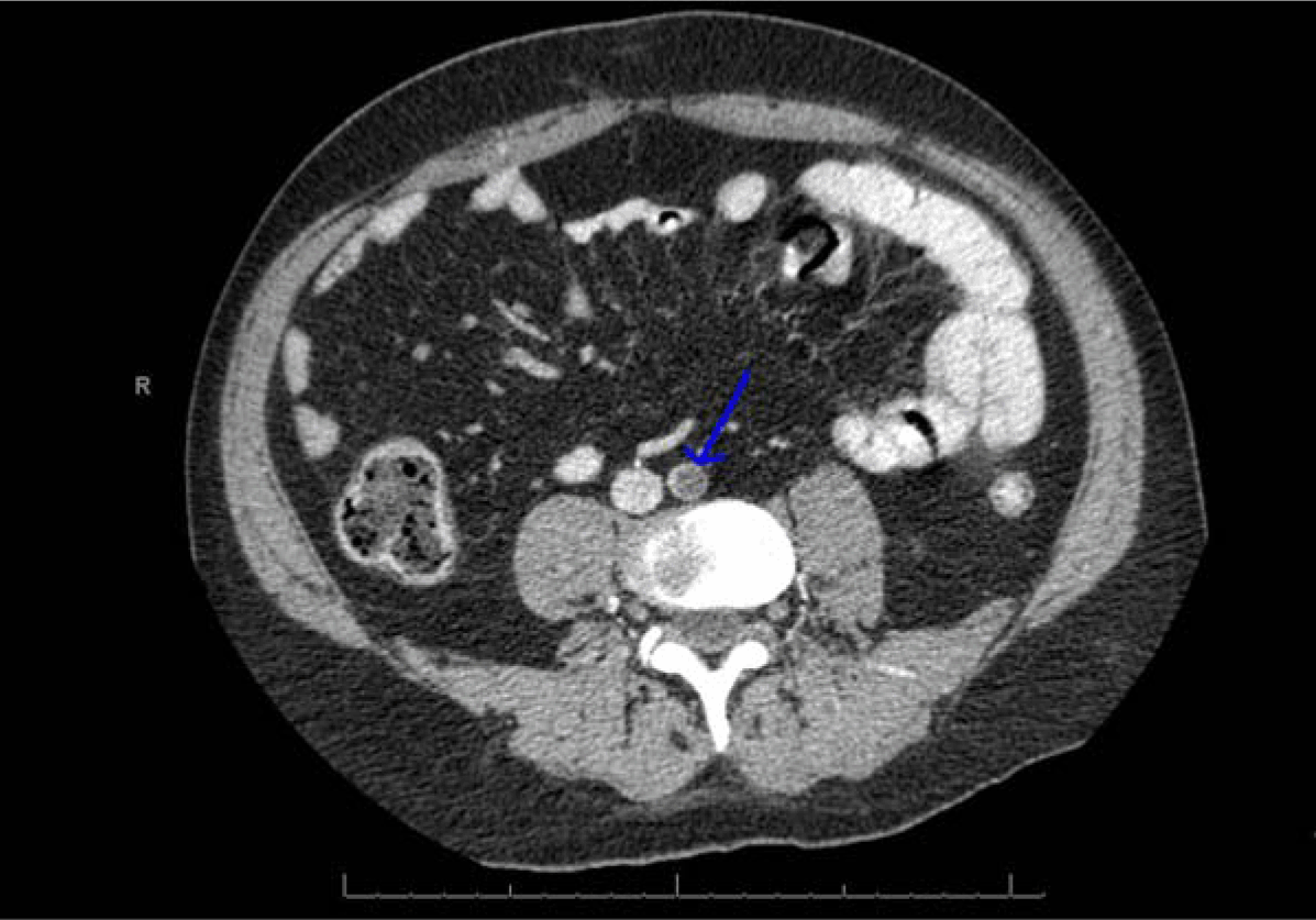
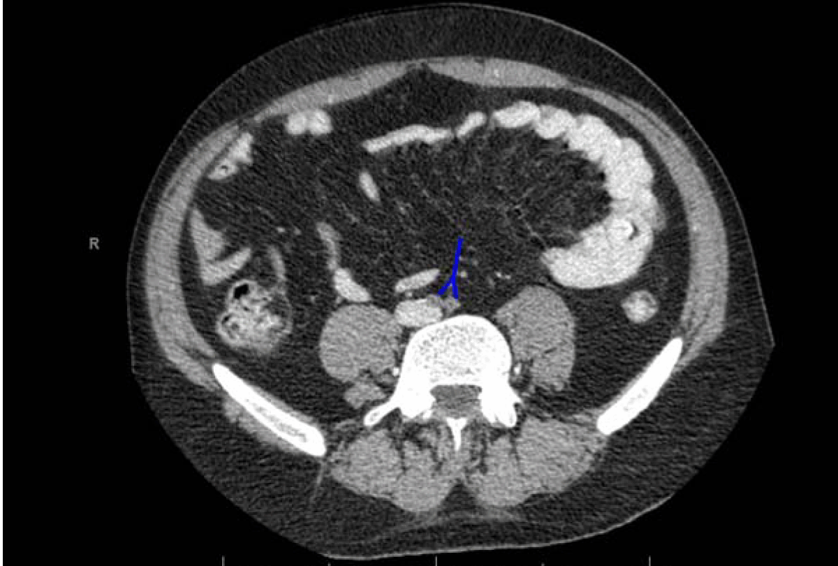
A 32-year-old British male presented to our emergency department as a referral from an outlying hospital. He was complaining of severe back pain and bilateral leg weakness. The onset was sudden and started an hour prior to arrival at the referring hospital. Associated symptoms included chest pain, bilateral upper and lower limb tingling, numbness and weakness. Further questioning also revealed a history of impotence for the past few months. Past medical and surgical history was significant for tetralogy of Fallot that was surgically treated at the age of ten years. Vitals on arrival were blood pressure 63/34 mmHg, temperature 35°C, pulse 118 beats/minute, respiratory rate 18/minute and oxygen saturation 100% on room air. On initial examination the patient was observed to be in severe distress, pale and diaphoretic. Chest auscultation was positive for decreased breath sounds on right upper, middle and lower lobes. The patient was also found to have severe bilateral lower limb weakness (left 2/5), better than right (1/5), pale skin, loss of sensation, and no palpable pulses bilaterally below both femoral arteries. As the patient was showing signs of hypovolemic shock, he was fitted with two large bore IV cannula and started on multiple normal saline boluses and empirically received two units of packed red blood cells (subsequent hemoglobin level 17.5 g/L). He was also covered with a heating blanket to counter hypothermia. A quick, portable antero-posterior view chest X-ray demonstrated right side diffuse parenchymal consolidation with pleural effusion. Arterial blood gas results showed pH 7.056, PCO2 46.7 mmHg, PO2 91.1 mmHg, bicarbonate 12 mmol/L, base excess - 17.4 mmol/L, sodium 139.7 mmol/L, potassium 5.5 mmol/L, calcium 1.19 mmol/L, glucose 4.8 mmol/L, and lactate 10.3 mmol/L. Laboratory results revealed several abnormalities but were most remarkable for an elevated Troponin-I 0.25 ng/mL, B-type natriuretic peptide 1.008 pg/mL, creatinine 164 mmol/L and urea 8.8 mmol/L. Vascular surgery and cardiology services were consulted to evaluate possibilities of aortic dissection or a vaso-occlusive thrombus. Following stabilization of the patient's blood pressure at 99/83 mmHg, he underwent emergent computed tomography (CT) scan with angiography. Imaging data demonstrated a saddle embolus in the distal aorta at the level of L4, extending into the right and left common iliac arteries (Figures 1 and 2), along with a suspicious embolus in the left renal artery at the mid-section causing an extensive renal infarction, and a splenic infarction at the ventral lower pole. There was also a massive cardiomegaly with dilated right ventricle. These findings confirmed the diagnosis of aortoiliac occlusion. The patient was immediately transferred to the operating theatre by the vascular surgeons, he underwent an embolectomy. The patient had cardiac arrest on the operating table with a cardiac rhythm of pulseless electrical activity (PEA) while waiting for an intensive care unit (ICU) bed to be prepared. Cardiopulmonary resuscitation (CPR) was performed with successful achievement of return of spontaneous circulation (ROSC). The patient was transferred to the ICU on light sedation. He was responding to pain. A bedside echocardiogram was performed by the attending cardiologist that demonstrated severe biventricular dysfunction and pulmonary hypertension. The patient unfortunately arrested again in the ICU two days later while on three inotropes. Cardiopulmonary resuscitation was unsuccessful on this occasion and the patient was pronounced dead. | ||||||
| ||||||
|
| ||||||
|
Discussion
| ||||||
|
Rene Leriche in 1923 described occlusion of the distal aorta and iliac arteries, which includes both acute and chronic forms of the disease [1] [3]. Acute Leriche syndrome most commonly presents with symptoms of acute limb ischemia. Clinical symptoms are dependent upon the level of arterial occlusion [3]. Isolated aortoiliac occlusive disease more commonly occurs in young female patients with a higher incidence of smoking and hypercholesterolemia as associated risk factors that portend a better prognosis. However, patients with a more multilevel pattern of the disease are commonly older males, and are more likely to have diabetes and hypertension as risk factors. These patients tend to have lower life expectancy [4][5]. This case report illustrates the importance of early identification of patients with relevant risk factors and symptoms of aortic occlusion. An important early warning sign in this case was absence of distal pulses coupled with back pain. There are several similar reported cases which ended in varying outcomes depending on rapidity of diagnosis and treatment initiation [2] [3] [6] [7]. Patients classically present with a history of the following triad of symptoms: claudication, impotence and decreased lower limb pulses. Alternatively, patients may present with a variety of symptoms like leg pain or weakness, loss of sensation in the lower limb and back pain as in this case [3]. Neurologic findings may initially lateralize, delaying the imaging needed to make this diagnosis [6] . Risk factors include hypertension, diabetes mellitus, hyperlipidemia and smoking [7]. Management in this case is usually surgical, aimed at relieving the symptoms and prevention of thrombus propagation. Recent reports advocate the use of ultrasonography for fast detection of aortic occlusion in the emergency department. It is a quick, inexpensive and reliable method for trained providers and can save a lot of time in delivering the definitive treatment to the patient [8]. | ||||||
|
Conclusion
| ||||||
|
This case of an acute presentation of Leriche syndrome highlights the critical need for early diagnosis and risk factor assessment in determining the outcome. A high index of suspicion is required. Bedsides, ultrasonography may prove to be an effective tool in the diagnosis of Leriche syndrome in the emergency department that can be used to screen critical patients presenting with unique symptoms, saving valuable time and shortening the interval until the patient receives definitive care. | ||||||
|
References
| ||||||
| ||||||

|
[HTML Abstract]
[PDF Full Text]
|
|
Author Contributions
Omar Sherif Askar – Conception and design, Acquisition of data, Analysis and interpretation of data, Drafting the article, Critical revision of the article, Final approval of the version to be published Abdel H Noureldin – Conception and design, Acquisition of data, Analysis and interpretation of data, Drafting the article, Critical revision of the article, Final approval of the version to be published Satish Chandrasekhar Nair – Conception and design, Acquisition of data, Analysis and interpretation of data, Drafting the article, Critical revision of the article, Final approval of the version to be published |
|
Guarantor of submission
The corresponding author is the guarantor of submission. |
|
Source of support
None |
|
Conflict of interest
Authors declare no conflict of interest. |
|
Copyright
© 2014 Omar Sherif Askar et al. This article is distributed under the terms of Creative Commons Attribution License License which permits unrestricted use, distribution and reproduction in any medium provided the original author(s) and original publisher are properly credited. Please see the copyright policy on the journal website for more information. |
|
|
|
About The Authors
| |||
| |||
| |||
| |||